

Welcome
Dive into the Exciting World of Protein Dynamics Beyond Static Structures!
DISCOVER THE THRILLING SCIENCE OF ENSEMBLE ENGINEERING!


Proteins operate as dynamic ensembles rather than fixed structures,
navigating evolving landscapes shaped by transient intermediate states
Unlocking protein ensembles and transient intermediates to power next-generation ligands and diagnostics.
Building robust, stable enzymes through ancestral protein design informed by ensemble landscapes.


The task is not to see what has never been seen before but to think what's never been thought before
- Erwing Schrodinger




Core Technical Expertise for Ensemble Analysis
Data from each of these tools are integrated in a novel way to provide information on high-energy intermediates & characterize ensembles
Protein Folding
Protein folding using wet-lab approaches, including thermal and chemical denaturation studies, monitored by:

-
Intrinsic fluorescence spectroscopy
-
Circular dichroism spectroscopy
-
Multi-angle light scattering (MALS)
-
Differential scanning fluorimetry (DSF)
-
Complex folding models derivation and fitting
-
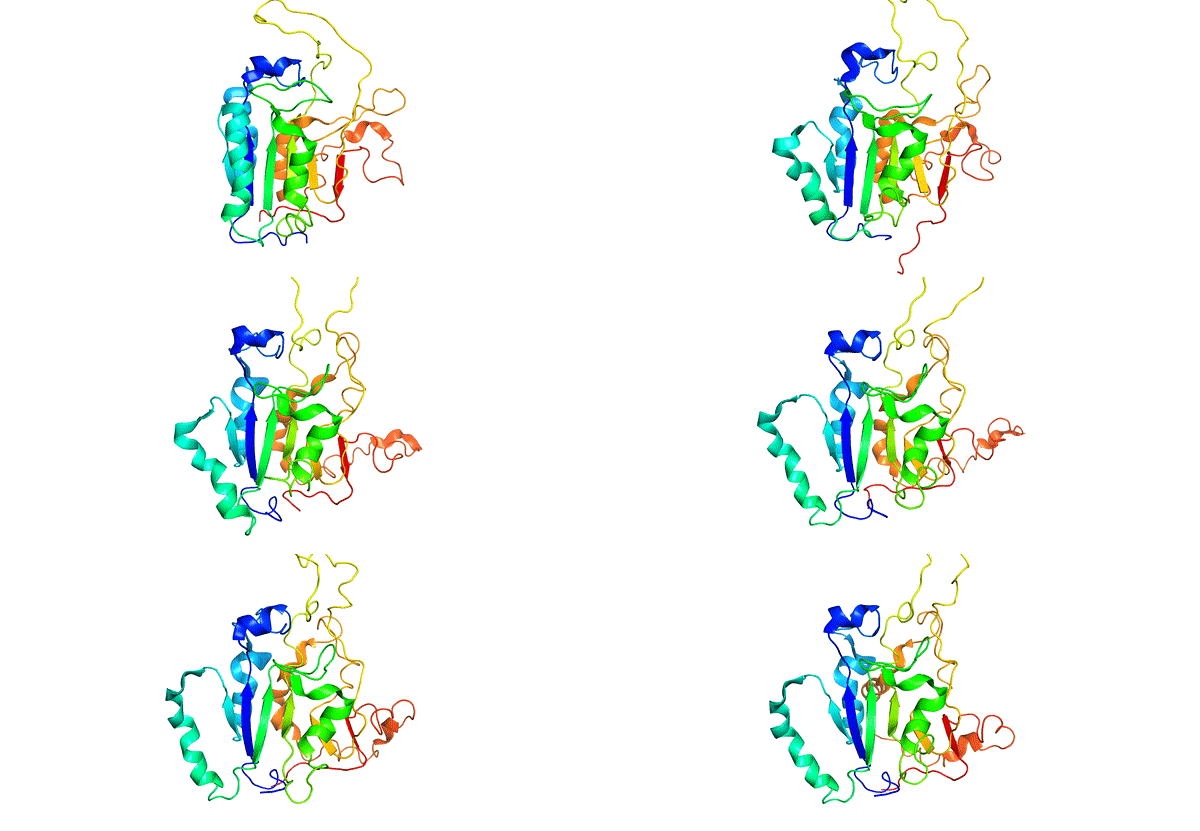
Conventional Simulations
-
Urea Simulations
-
Accelerated MD simualtion
-
Replica exchange simulation
-
Hamiltonian Replica exchange simualtions
-
SMOG simualtions
-
Umbrella Sampling
-
Metadynamics
NMR

-
N15 labelling
-
N15 C13 labelling
-
Proton NMR
-
2D HSQC and HNQC
-
Peak assignments and data analysis
-
Ligand binding analysis
TR-FRET

-
Coregulator binding affinity
-
EC50 from ligand binding
-
Complementary technique - Fluoresence Polarization for binding assays
Other Biohysical Studies

-
Bio-layer interferometery
-
DLS
-
High-throughput screening
-
FTIR Spectroscopy
-
ITC
-
Absorbance Spectroscopy
-
HPLC
-
Size exclusion chromatography
-
Nano DSF
-
SEC-MALS
-
Mass Photometery
Cell Stuides

-
HEK293T cells & 3T3L1 cells
-
Luciferase assay for in-vivo ligand binding
-
qPCR
-
Immunofluorescence
Mass Spectroscopy

-
MALDI-TOF
-
LCMS
-
Native Mass spectrometry
-
Proteolysis analysis
Crystallography

-
Crystal setup and screening
-
Mosquito nano-liter
-
Looping and data collection
Cloning

-
Plasmid mini prep and maxi prep
-
PCR
-
Site-directed mutagenesis
-
Gibson cloning
Ancestral Protein Reconstruction

-
Sequence retrieval from NCBI
-
Bulk sequence analysis on Jalview
-
Sequence Alignment
-
IQ-TREE tree generation
-
Marginal and Joint reconstruction with FAST-ML & GRASP suite
Other Expertise

-
Atomic Force Microscopy
-
Microfluidics
-
Python
-
Statistical Analysis
-
Jupyter Notebook analysis
-
HPC environment
-
Slurm scripts
-
Linux operating
Communication

-
Manuscript preparation
-
Presented poster and oral talks at a number international conference
-
Mentoring
-
Animations
-
Illustrations using adobe suite
-
Pymol & chimera
-
VMD


The Importance of Art in Molecular Biology
Art is essential to molecular biology because it can generate accurate and engaging visual aids for communication and comprehension. Scientific illustration and three-dimensional modeling can be used to illustrate complex systems and communicate discoveries to a larger audience. I am passionate about making artwork and cartoons to illustrate research findings.
This image illustrates key findings from my first study (click on the button below) showing how the structure of caspase-8 changes with pH. At neutral pH (first bubble), the protein is highly stable. As the pH becomes more acidic (middle bubble) or more basic,(bottom bubble) the structure is destabilized. The study highlights how different environments affect the balance of conformations in the protein ensemble, providing an ensemble picture of caspase-8.


Artwork featured on the cover of the Biochemical Journal
Pleased to see this artwork featured on the cover of the Biochemical Journal. The image illustrates how apoptotic caspases behave as dynamic energy landscapes rather than fixed structures. Instead of existing in a single shape, caspases transition between multiple states, from inactive to fully active, passing through higher-energy intermediate forms. The artwork highlights key differences between initiator and effector caspases and shows how evolution has shaped their energy landscapes to favor different functional states, such as monomers or dimers. These hidden intermediate states help explain how caspases are regulated and how they respond to cellular signals.
To read more about this work, please click on the buttom below


Animations to disseminate scientific information
Scientific animations are essential for disseminating scientific information because they provide a visual depiction of complicated concepts, make knowledge more accessible, interest the audience, show experiments and techniques, and are easily shareable across multiple platforms.
I am therefore compelled to make animations utilizing MD simulations, platforms, and Adobe apps to bring molecules to life and enlighten others about our discoveries.
Caspase activation cascade illustrated with adode animate
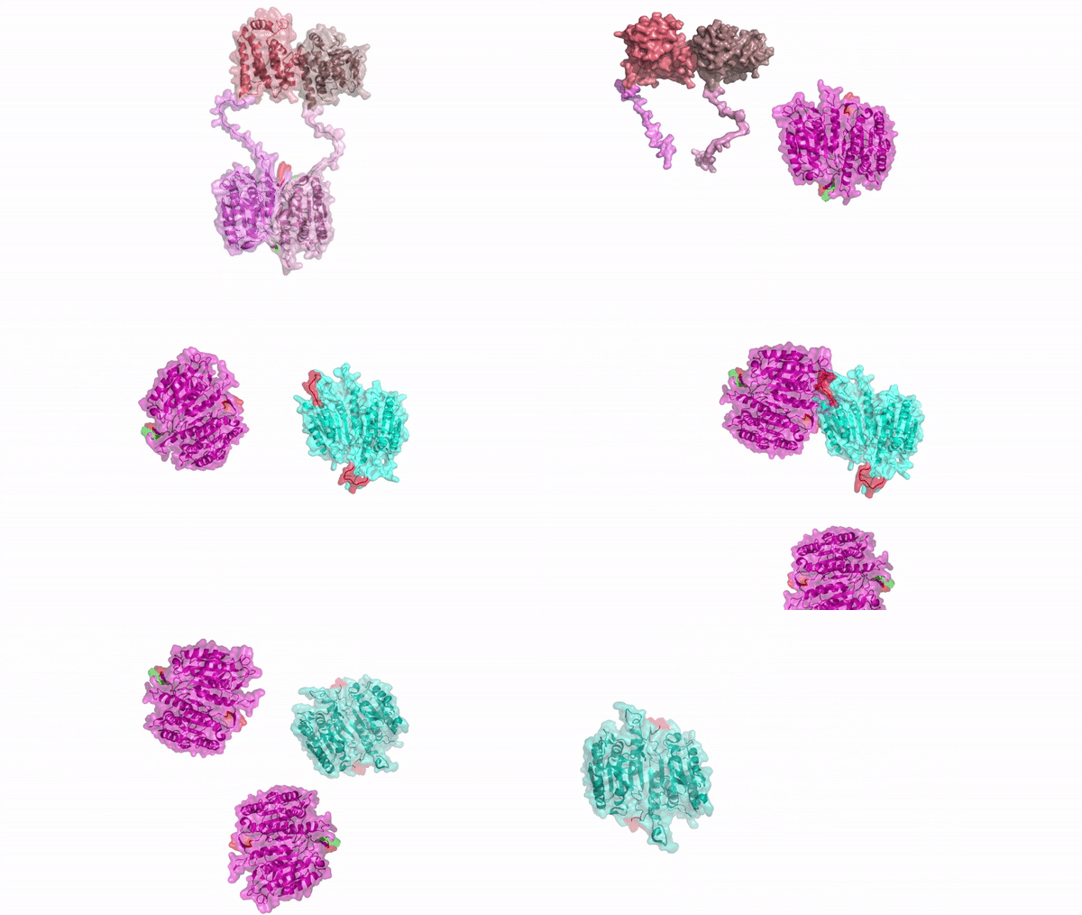
Purple: Caspase-8; Cyan: Caspase-3.
Caspase-8 functions as an initiator caspase, undergoing auto-processing to activate the apoptotic cascade. Activated caspase-8 then cleaves and activates the effector caspase-3, which executes downstream apoptotic processes.
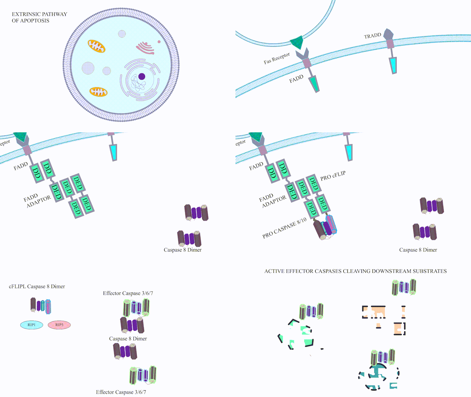
Cartoon animation of the Extrinsic pathway of apoptosis
Working Principle of Lanthan screen TR-FRET in PPARg LBD
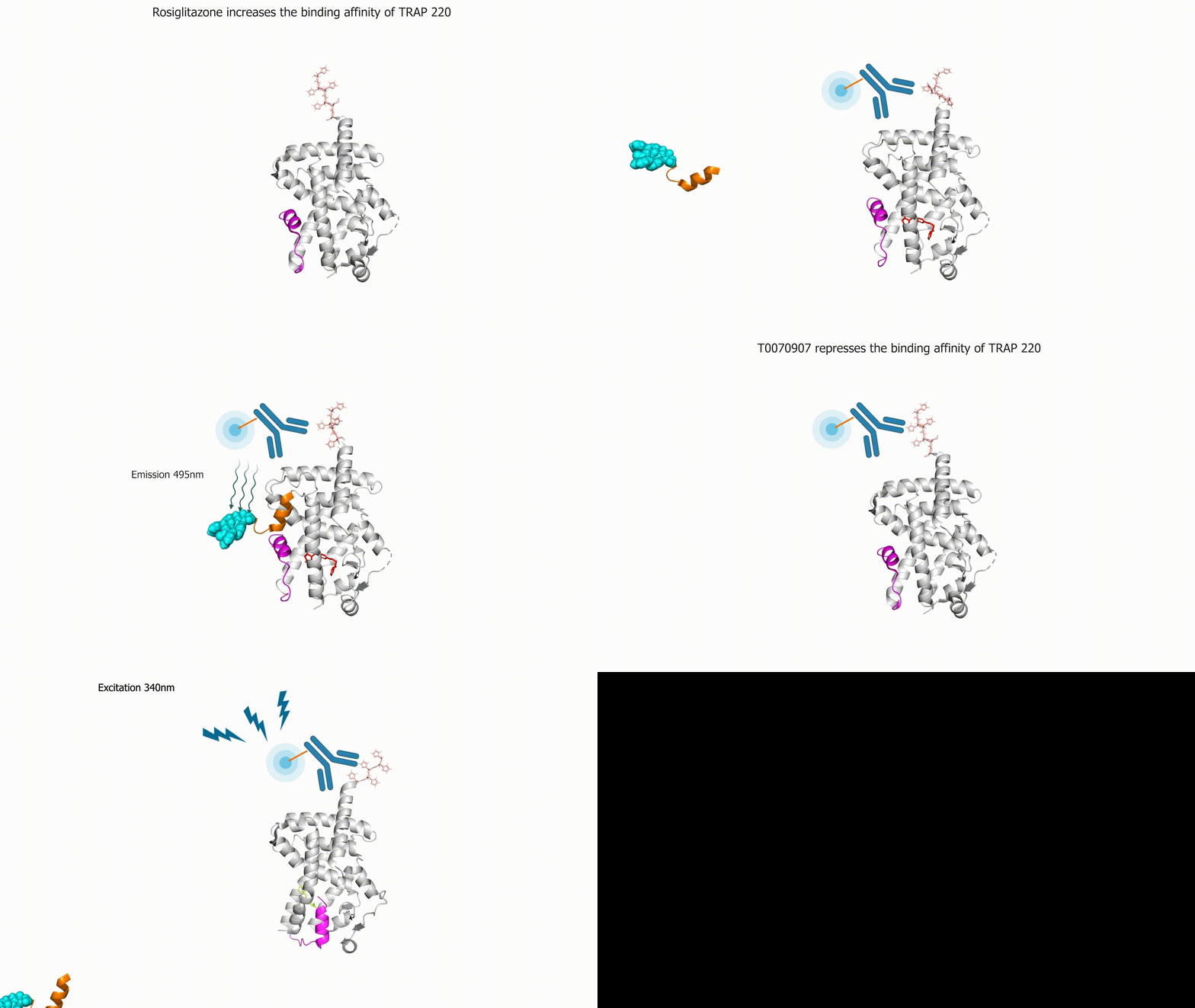
Refer to thermofischer website for technical details on this product
Publications
-
Nag, M. & Clark, A. C. Conserved folding landscape of monomeric initiator caspases. Journal of Biological Chemistry (2023).
https://doi.org/10.1016/j.jbc.2023.103075 -
Bibo-Verdugo, B., Joglekar, I., Nag, M., Ramirez, M. L., Snipas, S. J., Clark, A. C., Poreba, M., & Salvesen, G. S. Resurrection of an ancient inflammatory locus reveals switch to caspase-1 specificity on a caspase-4 scaffold. Journal of Biological Chemistry (2022), 298, 101931.
https://doi.org/10.1016/j.jbc.2022.101931 -
Joglekar, I., Nag, M., Diaz, D. A., Deo, A., & Clark, A. C. Evolution of the conformational ensemble and allosteric networks of apoptotic caspases in chordates. Biochemical Journal (2025), 482(15), 1029–1046.
https://doi.org/10.1042/BCJ20250001 -
Zorc, S. A., Munoz-Tello, P., O’Leary, T., Yu, X., Nag, M., Hondros, A. D., Hansel-Harris, A., Forli, S., Griffin, P. R., Kojetin, D. J., Roy, R. N., & Janiszewska, M. Structural insights into IMP2 dimerization and RNA binding. Journal of Structural Biology (2025).
https://www.sciencedirect.com/science/article/pii/S1047847725000826 -
Laughlin, Z. T., Arifova, L., Munoz-Tello, P., Yu, X., Nag, M., Dong, J., Harp, J. M., Zhu, D., Kamenecka, T. M., & Kojetin, D. J. Structural basis of PPARγ-mediated transcriptional repression by the covalent inverse agonist FX-909. Journal of Medicinal Chemistry (2025).
https://pubs.acs.org/doi/full/10.1021/acs.jmedchem.5c01252 -
Arifova, L., MacTavish, B. S., Laughlin, Z. T., Nag, M., Shang, J., Li, M.-H., Yu, X., Zhu, D., Kamenekca, T. M., & Kojetin, D. J. Shifting the PPARγ conformational ensemble towards a transcriptionally repressive state improves covalent inhibitor efficacy. eLife (2026). https://elifesciences.org/articles/106697
YouTube videos
This section contains links to educational content on my YouTube page
Will be adding more content soon addressing research findings